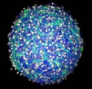
 A tiny cell culture; a tiny brain. A tiny cell culture; a tiny brain. by Kyu Bin Kwon ’18 Just imagine: small brains. More specifically, small cell cultures of a three-dimensional spheroid central nervous system (CNS), made in Petri dishes, about a third of a millimeter in diameter. Feeling excited yet? Neuroscientists sure are, and researchers at Brown have found ways to make these supplies of mini-brains even cheaper and faster in a new study. These tiny brain models provide so much potential for the studies of CNS function, disease and therapeutics. Barry Connors, chair of the Department of Neuroscience, attributes the novelty of mini-brains to their depth and shape. 2D cell cultures that are widely used in present works are limited in that there are only lateral intercellular connections; alternatively, 3D models successfully mimic the complexity of microenvironmental cues in the brain and are “more relevant to the in vivo [living] scenario,” according to graduate student and co-author of the study, Molly Boutin. Connors wrote that the brains might be “useful for studying the movement of molecules like drugs, nutrients and transmitter molecules through the complex matrix of brain cells,” and added that “they could be a good place to study the molecular signals that allow neurons to develop and hook up into the sort of organized circuits that comprise real brains.” Diane Hoffman-Kim, professor of medical science and engineering and the corresponding author of the study, further predicts that the mini-brains may reduce the amount of animal testing as the effectiveness of drug treatments can be tested prior to clinical trials. Brown biologists and bioengineers outline the originality in the newfound method of mini-brain production in their paper produced in the journal Tissue Engineering: Part C. “The materials are easy to get and the mini-brains are simple to make,” as co-author of the paper Yu-Ting Dingle summarized. The method only requires postnatal cultures as opposed to progenitor or embryonic cell isolation. The mini-brains can be manufactured within 2 weeks, with electrically active neurons, glia, and cell-synthesized matrix. Turns out that mini-brains really are simple to make: the researchers extracted a part of rodent brain, broke it into single cells, and put them into unique three-dimensional Petri dish that aren’t used for most cell cultures. Once the single cells fall into the molds of the Petri dish, they self-assemble to form little spheres. The ease of production, coupled with the low cost of 25 cents per mini-brain, would allow this technology to be implemented in all labs—it could enable and catalyze all sorts of research. This new method endows mini-brains with several important properties. In addition to its 3D structure, these cell cultures have similar density to natural rodent brains and can live for at least a month. There is diversity in cell types, as the cultures have both inhibitory and excitatory neurons and a variety of neural support cells called glia. Also, the mini-brain cells produce their own extracellular matrix to produce tissue with the same “squishiness”—as Hoffman-Kim put it—as natural tissue. Since the manufacture of mini-brains simply involves injection of cell solution in agarose hydrogels microwells, the procedure does not rely on foreign materials like collagen. And don’t worry; there shouldn’t be ethical concerns about reproducing brains or nights lost pondering over whether a world domination led by sentient mini-brains looms in the future, etcetera. The mini-brains, while imitating most of the natural brain’s properties, cannot perform cogitation. The day of neuroscientifically-inclined-American-Horror-Story-meets-reality is yet to come; instead, the mini-brains have drawn the curtains to a whole world of new research waiting to take place—the start of a much cheerier and exciting scenario.
0 Comments
Leave a Reply. |
